
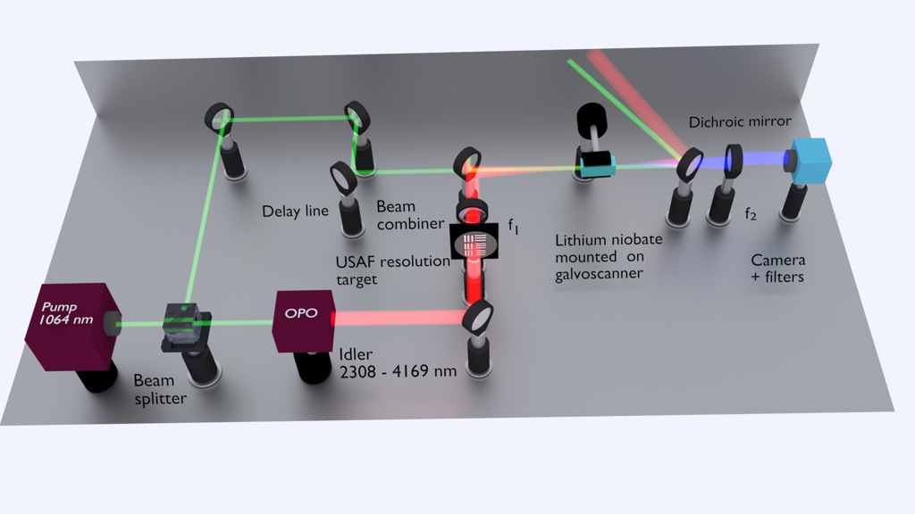
Illustration of the setup for upconversion-based mid-infrared hyperspectral imaging. [Image: Chaitanya Kumar, ICFO and Saher Junaid, DTU Fotonik] [Enlarge image]
Mid-infrared (mid-IR) spectroscopy is a useful tool for chemical analysis, providing detailed information about the molecular makeup and dynamic processes of a sample. However, its widespread use is limited by the expensive equipment and complex software needed to transfer data in the mid-IR spectral range into useful images.
A research team from Europe now says it has developed a simplified technique for producing mid-IR images for hyperspectral imaging (HSI) by upconverting the mid-IR signal to the easier-to-process near-IR region (Optica, doi: 10.1364/OPTICA.6.000702). The new system uses a single pulsed light-source, a crystal-based upconversion system and a standard CCD camera to create video-rate, information-dense images that require minimal post-production.
In proof-of-principle demonstrations with esophageal biopsies, images created using the mid-IR HSI method clearly delineated cancerous tissue from healthy tissue. One of the paper’s authors, Peter Tidemand-Lichtenberg from DTU Fotonik, Denmark, said in an OSA press release that this imaging approach “could be combined with machine learning to enable faster, and possibly more objective, medical diagnoses based on chemical signatures without the need for [traditional histology] staining.”
Translating mid-IR to near-IR
Mid-IR HSI is a useful technology for a variety of applications—ranging from biomedical imaging to atmospheric and geologic monitoring—because wavelengths in the mid-IR range can relay molecular-level information. For biomedical imaging specifically, a mid-IR HSI system can reveal complex microscopic structures without traditional sample preparation and staining, potentially making it a valuable screening tool for cancer and other diseases.
Unfortunately, the system most commonly used for mid-IR HSI, called Fourier transform IR (FTIR), relies on special light sources and cameras, as well as expensive detectors that require cryogenic cooling. These FTIR systems also generate a significant amount of data, which necessitate complex post-processing software to produce readable images. Tidemand-Lichtenberg and his colleagues’ solution to the FTIR problem uses upconversion to simplify mid-IR imaging.
While frequency upconversion is not a new technology, the researchers are using it in a new way: Instead of merely changing a laser’s wavelength, they are upconverting entire mid-IR images to near-IR without losing image quality or spatial data. In addition, they can use near-IR equipment for the imaging system, which is more readily available and affordable than the mid-IR counterparts.
Upconversion star
![]()
Computer-assisted biopsy classification of a tissue sample in one of the authors’ proof-of-principle demonstrations. [Image: S. Junaid et al., Optica, doi: 10.1364/OPTICA.6.000702]
The star of the new mid-IR system is an optical parametric oscillator (OPO) that produces 20-picosecond pulses tunable from 2.3 µm to 4.0 µm. The OPO uses 10 W of a 15-W Yb-fiber laser. The remaining 5-W beam is split off before the OPO and serves as the pump-field for the upconversion process.
After the pulsed OPO beam passes through the imaging sample, it is combined with the pump-field beam and then spatially and temporally overlapped in a lithium-niobite crystal. The crystal acts as the nonlinear medium for the upconversion process, shifting the mid-IR beam signals to near-IR. The upconverted near-IR signals are then captured by a standard CCD camera. The synchronized pulses from the illumination source create images with better signal-to-noise ratios, and therefore require less postprocessing.
Putting the system to the test
The researchers report that the system could capture 40 mid-IR images per second—which is fast enough for video—with a 35-µm spatial resolution and a 10-mm-diameter field-of-view (FoV). (The FoV was increased by a factor of five by rotating the crystal one degree.) These results matched the team’s theoretical calculations for the mid-IR HSI system.
In additional proof-of-principle demos, the researchers used their mid-IR HSI and computer algorithms to distinguish healthy and cancerous tissues from esophageal biopsies. With cancer and other diseases, a tissue’s molecular composition changes as the illness progresses. These changes can be detected by capturing mid-IR hyperspectral images from tissue biopsies and comparing them to known spectral signatures of cancerous and healthy tissues. And because these spectral signatures are quantifiable, they can, in theory, be detected by a computer.
Results from these experiments showed, according to the team, that the imaging system could capture enough hyperspectral data from the biopsies to identify cancerous and healthy tissues as well as standard histopathology techniques which require a pathologist’s evaluation. This “objective rather than subjective” approach to histology could, say the researchers, someday greatly improve disease detection and tracking.
In addition to DTU Fotonik, team members include scientists from ICFO–The Institute of Photonics Sciences in Spain and Exeter University and Gloucestershire Hospitals NHS Foundation Trust, both in the U.K.