
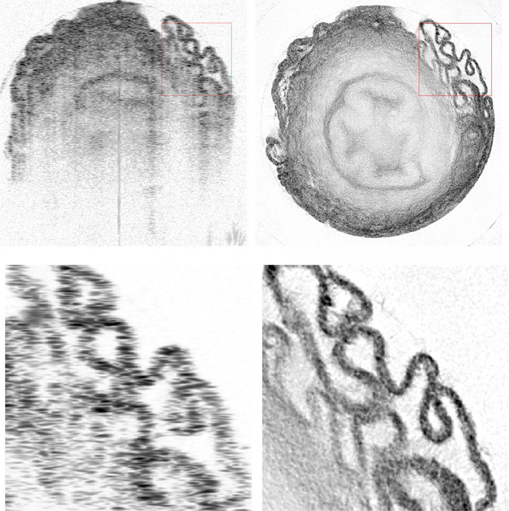
Image of a sample of mouse vas deferens tissue, from a normal optical coherence tomography (OCT) scan (left) and using a technique, optical coherence refraction technology (OCRT), that reportedly substantially boosts lateral resolution (right). [Image: Kevin Zhou, Duke University] [Enlarge image]
Optical coherence tomography (OCT) has become a staple of the ophthalmologist’s office, revolutionizing retinal imaging by providing cross-sectional images of retinal tissue at sub-micron axial, or depth, resolutions. But there’s historically been a trade-off, with users forced to accept relatively low lateral resolution for the sake of deep penetration and crisp imaging in the axial direction. That has made the technique great for tissues and structures that consist of relatively flat layers—but less practical when any lateral complexity enters the mix.
Now, a research team from Duke University, headed by OSA Fellow Joseph Izatt, has put computational-imaging techniques to work to offer a solution (Nat. Photon., doi: 10.1038/s41566-019-0508-1). The approach the researchers have unveiled, which they call optical coherence refraction tomography (OCRT), reportedly can boost the lateral resolution of OCT as much as threefold, to the micron scale, making it comparable to OCT’s axial resolution. The team believes that the advance could set up OCT for a wider range of applications not only in the ophthalmology clinic, but in imaging diseases and disorders in other biological tissues.
The anisotropy problem
OCT works by taking a series of 1-D laser interferometric scans, or “A-scans,” of layered tissue such as the retina. Those scans—effectively 1-D optical echograms showing different tissue layers at depth—are then stitched together to form a 2-D, cross-sectional image, the “B-scan.” Since its invention in the early 1990s, the technique has seen numerous improvements in both laser sources and computational techniques, and has expanded from use in ophthalmology to other areas in both medicine and industry.
Impressive as OCT’s success has been, however, the images it creates have remained stubbornly anisotroptic, owing to the tension between a large axial depth-of-field, which requires a relatively large laser beam diameter, and lateral resolution, which requires keeping the beam waist tight. Most practitioners have thus accepted lateral resolutions on the order of 10 microns or even coarser, for the sake of imaging tissues to depths of a millimeter or more at micron-scale axial resolutions.
That anisotropic view, the authors of the new study point out, can prevent OCT from picking up potentially meaningful ultrafine structures if they happen to be oriented obliquely to the optical axis. Some previous approaches attempting to overcome the problem and boost lateral resolution, specifically using holography, have worked—but only if the sample and imager remain perfectly still during the entire process. That is, Izatt noted in a press release accompanying the research, “very difficult to achieve in living tissues because they live, breathe, flow and change.”
Mapping index variations
Izatt and his grad student (and first author on the new study), Kevin Zhou, wanted to see if they could get to a more practical system that would enable an isotropic OCT view. To get there, they looked at the experience of an analogous technology—computerized tomography, or CT, scanning. In CT scanning, lateral resolution can be improved by taking projections in multiple different directions and algorithmically reconstructing the image from those multiple projections.
The researchers reasoned that something similar might be done to boost lateral resolution in OCT, by taking a series of B-scans at different angles. But to make it work, they needed to compensate for variations in refractive index in the tissue, which might otherwise gum up the result of the multi-angle approach.
To overcome that issue, Zhou, working with another Duke professor, biomedical engineer Sina Farsiu, developed a forward-modeling approach, based on ray tracing, that could solve iteratively for those variations to develop a refractive-index map of the sample, even as the multiple B-scans were being registered. The team then wrote up routines to implement the model using TensorFlow, a popular deep-learning library provided by Google.
From mouse organs to human corneas
With the theory and the code in hand, the team tested out the process—first on samples consisting of polystyrene beads, and then on animal tissues including a variety of mouse organs and donated human corneas. Their setup used a commercial spectral-domain OCT system and a custom-built, rotating sample stage, with the stage motions and data acquisition automated using Python scripts. The OCRT imaging resulted in striking additional detail owing to the technique’s improved lateral resolution and decreased speckle noise.
The technique still has some practical issues to address. An obvious one is that the initial experiments involved bits of ex vivo tissue, twisted and turned on a rotating stage with a fixed light source—fine for a sliver of mouse organ, but not practical for use on live patients in the clinic. The researchers believe that this issue could be addressed through geometries that use angularly scanning probes rather than sample rotation, coupled with “subsampling” strategies that would allow good results while reducing the number of B-scan angles required. Zhou is reportedly already at work on such strategies for one application, corneal imaging.
Izatt believes that—as these issues and some others, such as a relatively long data acquisition times in the initial proof of concept, are overcome—the technique, and other improvements, could take OCT well outside of its traditional home in the ophthalmic clinic.
“OCT has already revolutionized ophthalmic diagnostics by advancing noninvasive microscopic imaging of the living human retina,” he said in a press release. “We believe that with further advances such as OCRT, the high impact of this technology may be extended not only to additional ophthalmic diagnostics, but to imaging of pathologies in tissues accessible by endoscopes, catheters, and bronchoscopes throughout the body.”