
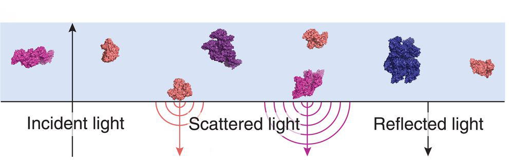
In the authors’ interferometric detection scheme, iSCAMS, the scattering signal scales with the polarizability, which is a function of the refractive index and proportional to the particle volume. That allows users to infer the mass of proteins from the scattering signal. [Image: From G. Young et al., Science 360, 423 (2018); reprinted with permission of AAAS] [Enlarge image]
European and U.S. researchers have reported a technique that uses light not only to observe individual biomolecules, but also to determine their mass (Science, doi: 10.1126/science.aar5839). Dubbed interferometric scattering mass spectrometry (iSCAMS), the method provides insight into molecular dynamics and could have applications in drug discovery and, ultimately, point-of-care diagnostics.
From imaging to mass measurement
In existing, fluorescence-based techniques for looking at biomolecular structures and interactions, molecules must first be labeled and excited, and then emission collected from them. Other, static methods involve averaging over many molecules in a sample, and thus can’t provide accurate spatiotemporal information or reflect the diversity in a sample. And state-of-the-art mass spectrometry works only in a vacuum, so it isn’t suitable for studying many biological systems in their living state.
The team behind the new research, led by University of Oxford chemists Justin Benesch and Philipp Kukura, sought a different approach—one flexible enough to look at small samples in solution, but without labelling and with improved spatiotemporal accuracy and resolution. They found a potential answer by leveraging interferometry. The researchers had, in fact, first used light scattering to image proteins back in 2014, and since then have improved the sensitivity of their technique to the point where they say it’s competitive with traditional fluorescence measurements.
The team also realized, though, that since the scattering signal scales with polarizability—which is a function of refractive index and is proportional to particle volume—its microscope should be sensitive to mass. More specifically, the researchers observed that there is very little variation (only around 1 percent) in the volumes of amino acids and the refractive indices of proteins. Since single amino acids can be considered as nano-objects, the team reasoned, the scattering signal should be proportional to the number of amino acids in a polypeptide, and thus to its mass.
From links to chains to amyloids
The group led by Benesch and Kukura, which also included other Oxford researchers and scientists from universities in Sweden, the United States, Germany and Switzerland, obtained high-quality images of single proteins diffusing from solution to bind with the interface between the microscope cover slip and solution. The signal-to-noise ratios were such that, by optimizing their data analysis, the scientists could precisely determine the scattering contrast for a single molecular binding event.
From there, the researchers obtained signatures for different oligomers—short macromolecule complexes consisting of a few simpler units—and their relative abundances. They repeated the experiments on eight different proteins to establish a linear relationship between mass and interferometric contrast, and confirmed the precision of the technique.
Once that was done, the researchers moved on to more complex systems. They were able to follow and model the evolution of various oligomeric species, and resolve changes in mass in both space and time; that enabled them, for example, to examine surface-catalyzed nucleation events that may eventually lead to the formation of amyloids, the proteins implicated in some neurodegenerative diseases (such as Parkinson’s). In other words, team co-leader Benesch suggested in a press release accompanying the work, the technique allows examination of questions such as whether molecules interact, how tightly, what the composition of a protein is, and how proteins grow or fall apart.
Broadly applicable
The relationship between volume, optical properties, and mass holds for molecules containing lipids and carbohydrates, as well as proteins, according to the team, so iSCAMS can be applied quite broadly. Indeed, the team finds that general applicability “tremendously exciting,” team co-leader Kukura said in a press release. Essentially, the researchers point out, every physiological and pathological process involves biomolecular interactions in solution—and mass is a universal property that reveals a lot about the molecule being investigated. The technique, Kukura says, allows users to see those properties and processes playing out in real time—using a compact, “shoebox-size” instrument that’s easy to operate.
The team is working on commercializing the technology and feels iSCAMS has the potential to “revolutionize how we study biomolecules and their interactions.”