
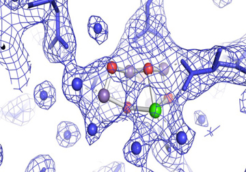
Water molecules are shown as blue spheres, manganese atoms are purple, calcium is green and the bridging oxygen atoms are in red. [Image: Jan Kern/Lawrence Berkeley National Laboratory]
Using a strong and fast X-ray free-electron laser, an international team of researchers has created high-resolution 3-D images of the water-splitting reaction in photosystem II—PS II, a protein complex involved in photosynthesis—under conditions that mimic nature (Nature, doi: 10.1038/nature20161). This work could bring scientists closer to understanding exactly how photosynthesis works, and could someday help develop cleaner energy sources driven by artificial photosynthesis.
The mysterious water-splitting catalyst
PS II is the first protein complex in photosynthesis. It collects and uses light energy to split water molecules into the oxygen we breathe, as well as hydrogen. While much is known about photosynthesis in general, this water-splitting reaction remains mysterious—at least on the atomic level. Previously, scientists used frozen PS II samples for imaging experiments, but this proved less than ideal, as freezing alters the protein’s structure and only provides information about its non-active “dark” state.
To help shed light on this mystery, the researchers used a ultrafast X-ray laser to investigate PS II’s oxygen-evolving metal catalyst (which consists of oxygen atoms bridging four manganese atoms with one calcium atom; see image), and how this catalyst uses light energy to split water molecules in a more natural environment.
Capturing photosystem II in action
To capture images of PS II’s water-splitting reaction, the team used the ultrafast X-ray free-electron laser from the Linac Coherent Light Source (LCLS) at SLAC National Accelerator Laboratory (USA). LCLS’ intense laser pulses last only 40 fs, which means the pulses can hit a room-temperature PS II sample and travel fast enough to deliver their data message—all before the sample is destroyed.
At SLAC, the researchers placed droplets of PS II crystals in solution on a moving tape and exposed the droplets to visible green light at room temperature in order to start the water-splitting reaction. Next, the samples were hit with laser pulses, which carried diffraction data to two different detectors in the LCLS, before the crystals were destroyed.
Software algorithms developed by the team translated the diffraction data into 3-D renderings of PS II in action at a spatial resolution of 2.25 angstroms. Since the X-rays destroyed the crystals, the researchers had to use hundreds of samples to gather enough data to construct images of each stage within the water-splitting reaction.
The team’s work not only produced high-resolution images of active PS II; it also provided unexpected insights into two steps in the four-step water-splitting process. The team was surprised, says co-principal investigator Vittal Yachandra of the University of California, Berkeley (USA), to find that “the two leading theories explaining the mechanisms for how the reaction proceeds are probably not correct.” The researchers plan to continue their work, with their ultimate goal being to “emulate what photosynthesis has been doing for about three billion years.”
This collaboration spanned the globe and includes team members from the Humboldt University of Berlin, Germany; Uppsala University and Umeå University, Sweden; Brookhaven National Laboratory, USA; SLAC National Accelerator Laboratory, USA; Stanford University, USA; University of California, Berkeley, USA; and the University of Oxford, U.K.