
A diagram showing how the 3-D architecture of a capillary bed can be reproduced in hydrogel using short-pulse lasers. Scale bars: 100 μm. Credit: M. Lütolf / EPFL
A team of researchers from Switzerland’s Ecole Polytechnique Fédérale de Lausanne (EPFL) has developed a short-pulse laser-based technique for carving 3-D tunnel-like microfluidic structures in cell-culture hydrogels (Adv. Mater., doi: 10.1002/adma.201601099). They say that that their technique has the potential to advance tissue engineering by creating biocompatible structures that can influence cell function and promote tissue formation.
An organ’s microenvironment is important because it serves as the delivery route for various cell-signaling molecules that control how cells interact with each other to support tissue health and function. However, most cell-culture and microfluidic techniques are conducted in 2-D. These flat structures—like Petri dishes and microchips—cannot replicate the real-life 3-D microenvironments that exist in the liver, kidneys, lungs, heart, etc.
To create a bio-friendly 3-D microenvironment, the researchers, led by EPFL’s Matthias Lütolf and Ph.D. student Nathalie Brandenberg, used a focalized short-pulsed laser to carve pathways and networks into hydrogel “scaffolds.” The short-pulsed laser, which is a common tool in cell biology, can also be used while the cells are growing in the hydrogel, allowing for real-time adjustment to the delivery of cell-signaling molecules.
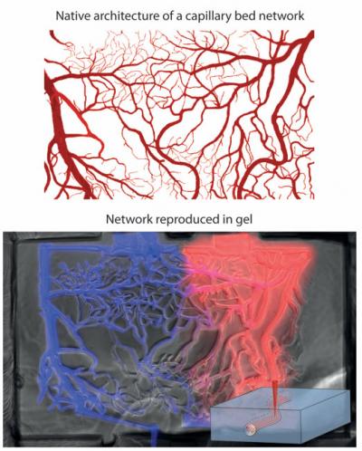
The EPFL team demonstrated their method by replicating a capillary bed using 3-D cell-culture hydrogel. First, they used a thresholded binary image of a capillary-bed photograph to generate a “mask” and path file that served as a guide for the laser. Once the micro-channel structure was carved out, the researchers connected the network to a pumping system to perfuse the hydrogel with different fluorescent molecules so that they could observe how the artificial capillary bed mimicked arteriovenous circulation (see image).
The authors believe their laser-based technique represents “an important step forward in the implementation of miniaturized, near-physiological cell and tissue culture platforms to answer currently unresolved questions in biomedical research as well as the ability to guide cellular self-organization to form functional tissues in vitro.”