
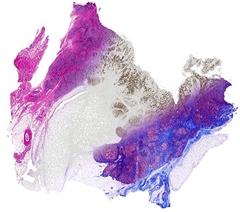
Breast tissue computationally stained using data from FT-IR spectroscopic imaging. Left to right: H&E stain (pink, blue), epithelial cell stain (brown) and Masson's trichrome (blue, red). Credit: R. Bhargava, University of Illinois
Spectroscopic data from a single infrared scan may give pathologists and researchers all the information they need to analyze a tissue sample, reducing the processing time and the cost associated with traditional tissue staining protocols (Technology, doi: 10.1142/S2339547815200010). The digital molecular pathology technique, developed by researchers and clinicians from the University of Illinois (USA), can quickly identify multiple markers without destroying the sample. This is important because tissue samples are often limited and histopathology results are important in time-dependent settings like drug response assays and disease diagnoses.
Traditional tissue staining protocols often require several steps and involve chemical dyes that can damage cells. The University of Illinois’ new digital technique uses Fourier transform infrared (FT-IR) spectroscopic imaging to produce spectra indicative of the cells’ chemical composition. The spectral data are processed with a computer algorithm that translates them into chemical stain patterns. Unlike physical staining, a pathologist can apply multiple computational stains to a single sample. Computational staining also eliminates imaging artifacts that occur when dyes aren’t applied evenly to a tissue sample.
The team, led by Rohit Bhargava, used human tissue samples to compare their computational stains with five common physical stains: hematoxylin and eosin (H&E), Masson’s trichrome stain, high molecular weight (HMW) cytokeratin, smooth muscle alpha actin and vimentin. Their new technique reliably replicated the physical stain patterns.
Using FT-IR spectroscopic imaging, “Any sample can be analyzed for desired stains without material cost, time or effort, while leaving precious tissue pristine for downstream analyses,” says Bhargava. The authors believe digital molecular pathology could enhance researchers and clinicians’ ability to make “molecularly informed decisions in the microscopic analyses of tissue.”