
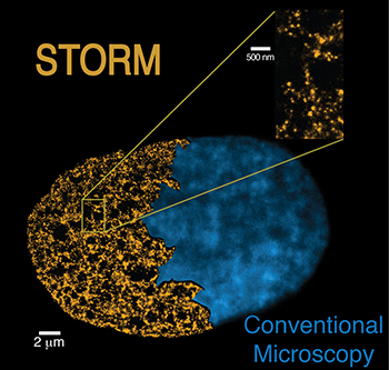
An image of the nucleus using the STORM technique (yellow) compared with conventional microscopy (blue). Credit: IFCO / CRG
A multidisciplinary team from the Institute of Photonic Sciences and the Centre for Genomic Regulation (ICFO, CRG; Spain) has used super-resolution microscopes to see and count nucleosomes—small packages of DNA that helps structure our chromosomes (Cell, doi: 10.1016/j.cell.2015.01.054). The researchers found that nucleosomes aren’t regularly spaced along DNA, as previously believed, and that their arrangement can indicate degree of stem cell pluripotency, i.e., the ability of a cell to give rise to other cell types in the body. These results could increase our understanding of stem cell differentiation and inform a standard method of quality control for stem cell use in medicine and research.
A nucleosome is made of a short segment of DNA wound around eight histone proteins. From a distance, a series of nucleosomes look like beads on a string of DNA; this complex is called chromatin. However, actually viewing nucleosome organization within chromatin in vivo has been impossible.
Physicists and biologists from ICFO and CRG combined efforts to leap over this imaging hurdle using a technique they call STORM (stochastic optical reconstruction microscopy). STORM uses quantitative super-resolution nanoscopy with computer simulations to non-invasively visualize and count nucleosomes along a chromatin fiber within a single nucleus. ICFO group leader Melike Lakadamyali says, “STORM overcomes the diffraction limit that normally restricts the spatial resolution of conventional microscopes and enables us to precisely define the chromatin fiber structure.”
They found that nucleosomes were grouped in “clutches” interspersed with lengths of nucleosome-sparse regions. After viewing and comparing chromatin organization in pluripotent and differentiated cells, they found that the median number of nucleosomes in the clutches and their level of compaction are specific to different cell types. Pluripotent stem cells had less dense clutches with fewer nucleosomes.
ICFO and CRG have filed a joint patent and hope to find commercial opportunities for their stem cell pluripotency classification technique to use in cell therapy or biomedical research.