
Occasionally, in paintings celebrated and obscure, hints of another version peek out from underneath the most recent layer of oil or acrylic—evidence of an earlier, unsuccessful draft subsequently covered up by the artist in the final rendering. The phenomenon is called pentimento, from the Italian word for “repentance,” a suggestion that the artist has changed his or her mind. Three recent studies have focused on optical and related techniques to help suss out, noninvasively, the layered history of paint and varnish on old and restored masterworks, and how ink affixes to paper in modern printing.
Looking over Rembrandt’s shoulder
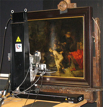
MA-XRF scanner trained on Susanna and the Elders, conservation studio of the Gemäldegalerie, Berlin. [Image: Matthias Alfeld]
In one of the studies (Appl. Phys. A, doi: 10.1007/s00339-015-9081-8), Matthias Alfeld of the University of Antwerp, Belgium, along with colleagues from Germany and the Netherlands, looked at a painting known to feature a large number of pentimenti: Susanna and the Elders, an oil-on-mahogany masterwork signed in 1647 by Rembrandt van Rijn. Alfeld and his colleagues wanted to determine which of several nondestructive methods of analyzing artworks best illuminated the history of the repainted canvas and, potentially, Rembrandt’s creative process.
The techniques the team studied included conventional X-ray radiography (XRR); neutron-activation autoradiography (NAAR), in which the painting is irradiated by a neutron beam and the beta radiation from the “activated” sample is allowed to expose photographic film; and scanning macro X-ray fluorescence (MA-XRF). Susanna had been subjected to both XRR (as early as 1931) and NAAR (in a 1994 study), but had not yet undergone MA-XRF. Comparing the methods had particular interest, since the relatively recently developed XRF can be deployed much faster than NAAR, which requires sequential exposures over a period of months.
Alfeld and colleagues began by subjecting the canvas to XRF over a weekend in the conservation studio. They then compared the new XRF results with earlier XRR and NAAR findings from the same picture. They discovered the XRF results were easiest to interpret (in addition to being relatively easy and fast to acquire), and were capable of revealing hidden features in pigments containing a range of elements, some of which are not visualized by NAAR.
They also found, however, that NAAR was better at probing deeper layers, being less subject to absorption by covering layers of paint. Moreover, both NAAR and XRR sported higher resolution than XRF, with the capability of discerning single brushstrokes and, potentially, revealing changes in technique. Thus, in the researchers’ view, while the speed and flexibility of XRF will make it increasingly common in future work, all three techniques will remain important in the art historian’s toolkit.
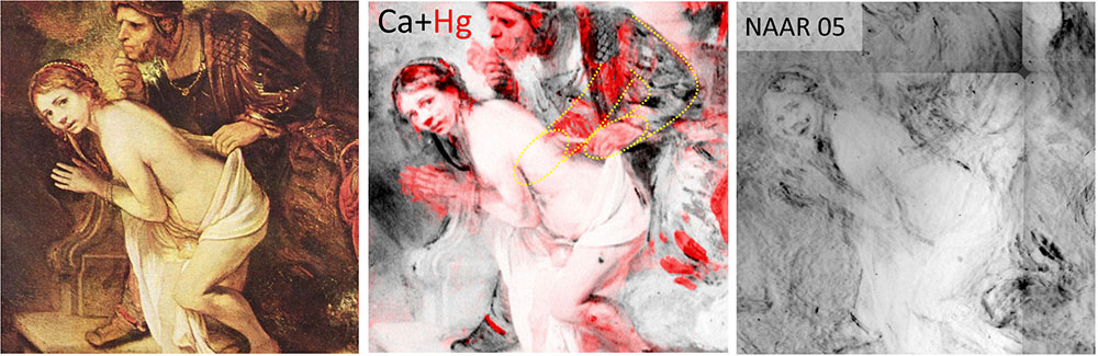
Left: Detail of Susanna and the first Elder, in normal light. Middle: XRF data on mercury content (red), a key component of the pigment used to depict the Elder's robe, reveals original and position of Elder's arm (yellow outline). Right: NAAR image from same segment reveals brushstroke details. [Image: Matthias Alfeld]
OCT for old masters
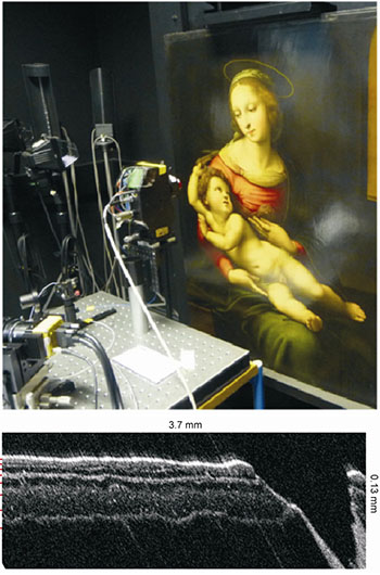
Top: FD-OCT setup, with pre-1600 copy of Raphael's Virgin and Child. Bottom: The OCT allows fine resolution of layers of conservator's varnish and painted strata. [Image: Cheung et al., Opt. Express, doi: 10.1364/OE.23.010145]
Another study, from a team led by Haida Liang at the University of Nottingham, U.K., devised a new instrument to bring a familiar optical imaging technique—Fourier-domain optical coherence tomography (FD-OCT)—to bear on a late-15th-century copy of Raphael’s “Bridgewater Madonna” (Opt. Express, doi: 10.1364/OE.23.010145).
Commercial OCT systems, long used in ophthalmological clinics to provide cross-sectional maps of structures in the eye, have already been applied to the problem of obtaining subsurface information from painted artworks. The relatively coarse depth resolution of the data from these systems (4 µm in paint or varnish), however, has limited their usefulness in art studies. Liang and colleagues attacked the problem by developing a new, ultra-high-resolution OCT setup that used a broadband laser source, which offered a wavelength palette that allowed scanning at a much finer depth resolution, to obtain information on features as small as 1.2 µm, across a thickness range as high as 1.5 mm.
The team applied the technique on a pre-1600 copy of Raphael’s The Virgin and Child (c. 1507) in London’s National Gallery, and reported that new setup could resolve very thin layers of restoration varnish on the canvas, at resolutions comparable to commonly used invasive techniques that require removal and destruction of a small cross-sectional sample of the painting. The team believes that the new technique, which they hope to make more widely available to art institutions, could ease the task of conservators seeking to understand the history of paint and varnish layers that have built up over years of art composition and restoration.
The topography of ink
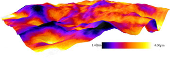
3-D visualization of the toner layer on top of coated paper. The dark blue areas show thin layers of toner, whereas the yellow shows thicker layers. [Image: Markko Myllys/University of Jyvaskyla]
A third recent study looked not at paint on wood, but on the more common phenomenon of ink on paper (J. Appl. Phys., doi: 10.1063/1.4916588). In the research, Finnish scientists led by Markko Myllys of the University of Jyvaskyla wanted to go beyond 2-D approaches to assessing ink on paper, such as scanning electron microscopy, to obtain a true 3-D view of the topography of ink—a perspective that could ultimately result in better, less expensive and more environmentally sensitive approaches to printing.
To get to that 3-D view, the scientists combined three techniques: X-ray microtomography, a technique analogous to a CT scan, to produce a 3-D picture of the underlying paper at around 1 µm spatial resolution; optical profilometry, which involved bouncing light off of the surface to obtain a surface profile of applied ink; and laser ablation, in which controlled amounts of ink were removed at various spots with laser energy, to allow a depth profile of the ink.
The result of this technique, according to the research team, was “a very realistic picture” of the interplay between ink or toner and its paper substrate, at a resolution that has been difficult to achieve previously. Among other things, the experiments highlighted the significant impact that microscopic surface roughness can have on the paper’s uptake of ink—considerably more significant, it turns out, than spatial chemical variations in a glossy paper’s finish. The team believes that the method will likely prove useful not only in analyzing the problem of print on paper but in studies of other thin films such as those in microelectronics, waterproof coatings, and solar panels.