
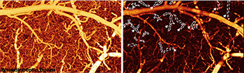
Left: Brain blood vessels in a healthy mouse brain (left), and a mouse brain exposed to cocaine.
A new 3-D imaging technique shows how cocaine disrupts cerebral blood flow (CBF) in the brains of mice. The laser-based technique uses optical coherence Doppler tomography (ODT) and was developed by researchers from Stony Brook University and the U.S. National Institutes of Health (Biomed. Opt. Express, doi: 10.1364/BOE.5.003217).
The team used ODT to direct laser light at blood cells moving through vessels. By measuring the refraction of the light that is bounced back, they were able to calculate CBF. The technique is similar to the Doppler effect experienced when a siren’s wail rises and falls in pitch as it approaches and recedes from the observer.
To test the method, scientists measured and imaged blood flow in the brains of mice that were given cocaine. After 30 days, they were able not only to see the expected dramatic drop in CBF, but for the first time, to witness cocaine-induced microischemia—a precursor to stroke. During “phantom flow” validation studies, the team tested the accuracy of their phase summation method for extending ODT range to allow for CBF measurement in capillaries. They reported a quantification accuracy of 91 percent.
A better understanding of how blood flows through the brain can help brain surgeons and neuroscientists study the effects of drugs or disease on the brain’s structure, metabolism and function. Stony Brook’s Yingtian Pan, team leader, states that their method could also be used to image capillaries in the eye and to monitor the growth of new blood vessels during tissue engineering.