
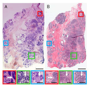
Breast tissue specimen imaged with NLM (a) and paraffin-embedded histology (b) with regions of cancer (red, blue and green boxes) adjacent to normal tissue. Credit: Proc. Nat. Acad. Sci. USA
A new microscopy technique can identify breast tumor margins during surgery, potentially enabling more accurate tumor removal in real-time and reducing the need for painful and costly additional surgeries. The U.S. research team, with members from MIT, Harvard Medical School and Thorlabs, Inc., used nonlinear microscopy (NLM) to image 179 fresh surgical specimens from 50 patients for tumor margin assessment. When compared with traditional paraffin or frozen-section analyses, NLM achieved 94 percent overall accuracy for identifying tumor borders for breast cancer in situ (Proc. Nat. Acad. Sci. USA, doi: 10.1073/pnas.1416955111).
The researchers performed NLM using a 100-fs tunable Ti:sapphire laser at 740 nm with a 76-MHz repetition rate, using a commercially available nonlinear microscope. They exploited NLM’s intrinsic and extrinsic contrast to obtain high-res images of morphological changes associated with breast cancer, like collagen reorganization, in breast tissue samples during surgery. They overcame limited field-of-view in high-magnification imaging with scanning and field mosaicking, resulting in subcellular resolution in square-centimeter-sized specimens. In practice, according to the authors, NLM findings would be confirmed with postoperative paraffin histopathology.
Breast cancer is the most common cancer and cause of cancer-related death in women worldwide. Traditional methods for identifying the margins of the cancer for surgery, however, have required tissue specimens to be embedded in paraffin, which can take up to 16 hours, or frozen, which is faster but sacrifices accuracy (sensitivities between 65 and 91 percent). As a result, about 40 percent of patients undergoing breast-conserving cancer surgery have to undergo additional procedures because postoperative pathology reports indicate that not all of the tumor was removed. The researchers hope that the NLM method—which could operate in real time and which sported high accuracy rates in their experiments—could forestall some of those repeat visits to the surgeon.
Currently, the scientists are redesigning their microscope to improve field-of-view and imaging speed. They also say that the NLM method could be used for lung, thyroid and head and neck cancer surgeries.