
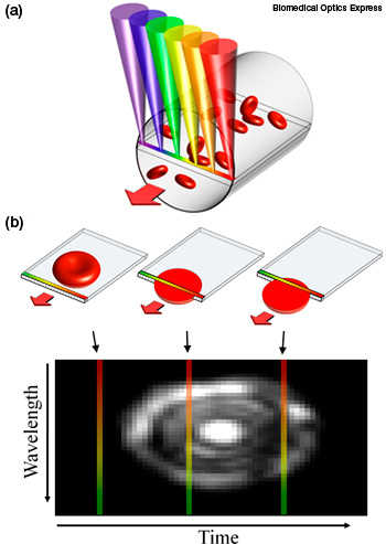
A new system for imaging blood cells in vivo uses spectrally encoded confocal microscopy. (a) A single line within a blood vessel is imaged with multiple colors of light that encode lateral positions. (b) A single cell crossing the spectral line produces a 2-D image with one axis encoded by wavelength and the other by time.
What if we could perform blood tests without drawing blood and without waiting for lab results? A new technology from Israel could noninvasively and continuously monitor patients’ blood flow during and after surgeries to detect internal bleeding or rapid inflammation. The device images blood flow through small blood vessels near the surface of the body using spectrally encoded confocal microscopy (SECM).
Lior Golan and others at Technion-Israel Institute of Technology and Rambam Medical Centre in Israel recently reported the first step in noninvasively imaging flowing blood cells (Biomed. Opt. Express 3, 1455). Using this method, researchers imaged the blood flowing through a vessel in the lower lip of a volunteer. They successfully measured the average diameter of the red and white blood cells and also calculated the percent volume of the different cell types—a key measurement for many medical diagnoses.
A microscope with a built-in green LED and a wide field-of-view is used to locate a small blood vessel near the skin’s surface. The cells within the vessel are then imaged by a rainbow of colors. The SEFC system uses broadband light from a fiber-coupled superluminescent diode array, split into different wavelengths by a transmission diffraction grating. The resulting line of light in a sequential rainbow of colors shines across the width of the blood vessel. The blood cells scatter light as they cross the line. The color of the scattered light carries spatial information that computer programs use to interpret the signal over time, resulting in 2-D images of the blood cells.
This first generation system will require more work—both to increase the penetration depth and to miniaturize the system. “Currently, the probe is a bench-top laboratory version about the size of a small shoebox,” says Golan. “We hope to have a thumb-size prototype within the next year.” A portable, rugged device could enable doctors in rural areas to screen many patients for common blood disorders without requiring lab access.