
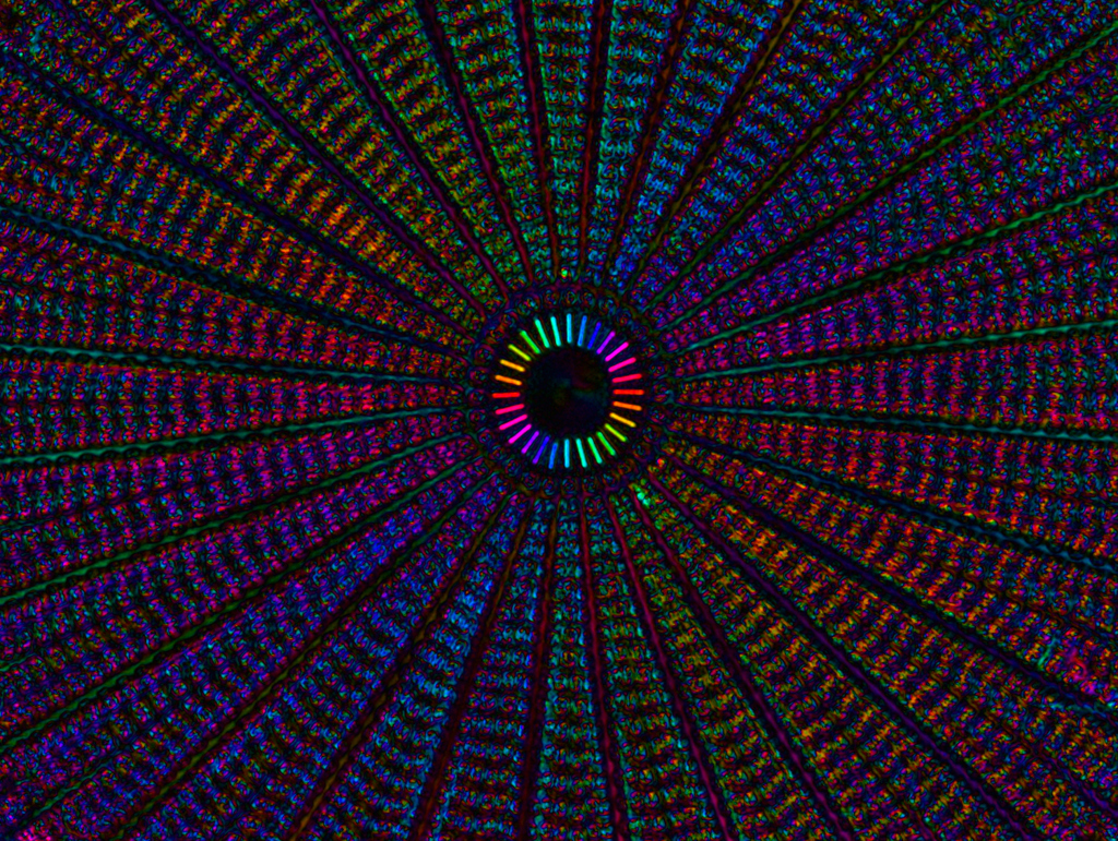
A Diatom's Details
This is a live image of the of diatom Arachnoidiscus. The picture shows the diatom's silicified cell wall, which forms a pillbox-like shell composed of overlapping halves that contain intricate and delicate markings. The picture was taken with the polychromatic polscope. An eye or camera can directly see the colored polarization image through the ocular with brightness corresponding to retardance and color corresponding to the slow axis azimuth. [Winner 2017 After Image photo contest.]
—Michael Shribak, Marine Biological Laboratory