
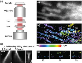
 (a) Schematic of the setup. (b) x-z views of the SB-PSF and the standard Gaussian PSF. (c) Conventional immunofluorescence images of mitochondria in BS-C-1 cells taken with the standard Gaussian PSF. (d) 3-D STORM images of the same areas taken with the SB-PSF. The z-position information is color-coded according to the color scale bar. White arrows indicate mitochondria that are undetectable in the conventional image (c). (e) Cross-sections along dashed lines (i), (ii) and (iii) in (d) show hollow outer-membrane structures of mitochondria. Scale bars: 500 nm (b), 1 µm (c-e).
Super-resolution fluorescence imaging techniques have overcome the optical diffraction limit (~λ/2) of conventional fluorescence microscopy, allowing visualization of biological structures and processes with near-molecular-scale resolution. Of these techniques, stochastic switching-based methods such as stochastic optical reconstruction microscopy (STORM) and (fluorescence) photoactivation localization microscopy ((F)PALM) rely on precise localization of the point spread functions (PSFs) of individual fluorophores. Researchers have developed various 3-D localization strategies by encoding the original Gaussian PSF with axial information.1 However, until recently we lacked a method that provided both isotropic 3-D resolution and large imaging depth.
In our new work, Airy beams provide a solution to the problem.2 As a nondiffracting waveform, Airy beams propagate over many Rayleigh lengths without appreciable diffraction, which considerably enhances the imaging depth.3 They also undergo lateral displacement as they propagate, resulting in a self-bending light path. Because of this curved trajectory, the axial position of an emitter could be determined from the lateral displacement of the beam.
We generated this self-bending PSF (SB-PSF) experimentally using a spatial light modulator. We then incorporated the SB-PSF into a STORM microscope and used it to demonstrate super-resolution imaging of various biological samples. Remarkably, the STORM images produced using the SB-PSF not only exhibited isotropic resolution of 20–30 nm in all three dimensions, but also imaged several micrometers into biological structures of microtubules and mitochondria in mammalian cells. This new approach offers significant improvement over previously reported 3-D localization methods.
We believe that the unique features of Airy beams will empower bioimaging in many applications, and lead to promising developments in optical design for biological research. For example, the self-healing effect that Airy beams exhibit after being obscured in scattering media has led to a recent development in Airy beam light-sheet microscopy.4
Researchers
Shu Jia and Xiaowei Zhuang, Harvard University, USA
References
1. B. Huang et al. Cell 143, 1047 (2010).
2. S. Jia et al. Nat. Photon. 8, 302 (2014).
3. G.A. Siviloglou et al. Phys. Rev. Lett. 99, 213901 (2007).
4. T. Vettenburg et al. Nat. Methods 11, 541 (2014).