
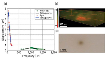
 (a) Frequency response spectrogram of agar and metal ball. (b) A 3-D ARF-OCE image excited at metal ball’s amplitude modulation frequency of 1,080 Hz. (c) Resonant elastography map of ball-agar sample.
Mechanical properties of living tissue can be crucial indicators of pathology, as many diseases can change those properties. Optical coherence elastography (OCE) offers a route to investigate biomechanical properties of tissues such as skin, muscle, cornea and blood vessels. But existing OCE techniques can have limited resolution and are difficult to adapt for in vivo imaging.
Two years ago, we developed a phase-resolved acoustic radiation force (ARF) OCE system, which uses an amplitude-modulated acoustic wave to apply dynamic pressure to vascular tissue and phase-resolved OCT to evaluate the tissue’s elastic properties.1 The system thus combines the high-speed excitation of ARF with the sub-micrometer detection sensitivity of phase-resolved OCT, and can achieve high-speed, high-sensitivity mapping of elastic properties of tissue.
ARF-OCE offers strong potential for clinical cardiovascular imaging—but it has also raised challenges for use in vivo. The reason is that, although the relative value of strain and Young’s modulus can be imaged with ARF-OCE, the absolute determination of these biomedical properties requires knowledge of ARF as applied to the tissue. We have developed a new technique that uses the mechanical resonant frequency to image and quantify tissue mechanical parameters, without advance knowledge of ARF parameters.2
Under small strains, the Young’s modulus of the tissue varies directly with the square of the resonant frequency.3 Measuring the ARF-OCE signal's frequency dependence thus allows direct determination of the biomechanical properties: With ARF as the excitation source, we can sweep different amplitude modulation frequencies and measure the frequency-dependent displacement. That, in turn, allows determination of the resonance frequency, which can be used to image and quantify the tissue's mechanical properties.
The spectral and resonant ARF-OCE method was first tested on an agar phantom, with a steel ball embedded inside. Very strong resonances emerged at 60 Hz for the agar and 1,080 Hz for the metal ball. When the agar phantom was imaged under the metal ball’s modulation frequency, the ball, as expected, showed up as a distinct vibration amplitude compared with the surrounding agar, yielding high contrast in the resonant elastography map. We confirmed the linear dependence of the resonant frequency on the square root of the Young’s modulus by measuring the frequency responses of silicone phantoms with varying Young’s moduli using the same method.2
Researchers
Zhongping Chen, Wenjuan Qi, Rui Li and Jiawen Li, University of California, Irvine, Calif., USA
Teng Ma, Kirk Shung and Qifa Zhou, University of Southern California, Los Angeles, Calif., USA
References
1. W. Qi et al. J. Biomed. Opt. 17, 110505 (2012).
2. W. Qi et al. Appl. Phys. Lett. 103, 103704 (2013).
3. C. Sun et al., J. Biomed. Opt. 16, 043001 (2011).