
Scatterings
Lighting Up the Inside of a Single Molecule
Researchers used luminescence spectroscopy and microscopy combined with STM imaging and tunneling spectroscopy to distinguish between vibronic couplings in orbitals in the same molecule.
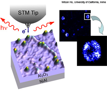 Bumps indicate molecules of magnesium porphine on an Al2O3 surface. (Inset) Zoom-in of light emission from a single molecule. The brightest regions correspond to transitions between two electron orbitals.
By combining scanning tunneling microscopy methods that use electrical and optical imaging, researchers could see optical transitions within a molecule with submolecular resolution. Researchers in Wilson Ho’s group at the University of California, Irvine, used STM-induced luminescence spectroscopy and microscopy combined with STM imaging and tunneling spectroscopy to distinguish between vibronic couplings in orbitals in the same molecule.(Chi Chen et al. Phys. Rev. Lett. 105, 217402, 2010.)
…Log in or become a member to view the full text of this article.
This article may be available for purchase via the search at Optica Publishing Group.
Optica Members get the full text of Optics & Photonics News, plus a variety of other member benefits.