
Scatterings
Two-Color STED Helps Unravel Protein Interactions
An advance in superresolution imaging will give scientists the ability to observe how proteins and other molecules interact in living cells in near real time.
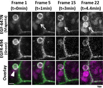 STED images using two fluorescent markers allows researchers to image the interactions between two proteins in living cells. In this case, the colors correspond to epidermal growth factor (EGF) in magenta and EGF receptors in green. The scale bar is 1 µm.
An advance in superresolution imaging will give scientists the ability to observe how proteins and other molecules interact in living cells in near real time. In a collaboration between researchers at Yale University and New England Biolabs Inc., Patrina A. Pellett and others recently have extended the ability of stimulated emission depletion (STED) microscopy to work using two colors (Biomed. Opt. Express, 2, 2364).
…Log in or become a member to view the full text of this article.
This article may be available for purchase via the search at Optica Publishing Group.
Optica Members get the full text of Optics & Photonics News, plus a variety of other member benefits.