
Scatterings
In-Vivo Images of Retina’s “Dark” Cells
A team at the University of Rochester (N.Y., U.S.A.) has taken the first images of this layer of so-called “dark cells” in a living retina.
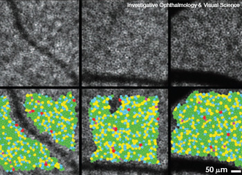 RPE cell mosaics (top) and corresponding Voronoi domain, or mathematical mapping, mosaics (bottom) at three locations on the retina of a human subject.
Retinal pigment epithelial (RPE) cells play a vital role in maintaining the health of the human retina, especially its photoreceptors (rods and cones). However, until recently, physicians had difficulty seeing individual RPE cells to assess them for potentially eyesight-robbing abnormalities.
…Log in or become a member to view the full text of this article.
This article may be available for purchase via the search at Optica Publishing Group.
Optica Members get the full text of Optics & Photonics News, plus a variety of other member benefits.