
Feature
Optical Biopsy with Confocal Microendoscopy
Using optical biopsy methods, doctors can potentially identify cancer and other conditions more accurately, in less time, and earlier in the course of disease than they can with standard screening techniques.
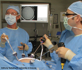 A confocal microendoscope is used to image the epithelial surface of an ovary in vivo.
Advanced medical imaging procedures such as computed tomography, ultrasound and magnetic resonance imaging have greatly improved physicians’ ability to detect cancer and other diseases. However, in order to make a definitive diagnosis, doctors often need to extract one or more tissue samples from a patient. This process is not only invasive, but can lead to high rates of misdiagnoses, especially when the disease is diffuse or at an early stage, because it is often difficult to know exactly where to extract tissue. In addition, tissue preparation and analysis are painstaking processes that are prone to human error. Moreover, extracted tissue that is fixed and processed for pathology is no longer alive—which means that we lose the ability to study active living processes that might shed more light on a patient’s state of health.
…Log in or become a member to view the full text of this article.
This article may be available for purchase via the search at Optica Publishing Group.
Optica Members get the full text of Optics & Photonics News, plus a variety of other member benefits.