
Scatterings
Supercharging Microscope Resolution
Researchers have refined a micro-scope that allows them to image features far smaller than the diffraction limit of the light used and examine how a protein behaves inside a living cell.
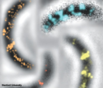 Four superresolution images show a structural protein (MreB) inside live Caulobacter bacteria cells. The red cell shows a ring at the division plane, where MreB is helping the cell divide. The other images show helical structures of MreB, which help maintain cell shape.
Researchers have refined a micro-scope that allows them to image features far smaller than the diffraction limit of the light used and examine how a protein behaves inside a living cell.
…Log in or become a member to view the full text of this article.
This article may be available for purchase via the search at Optica Publishing Group.
Optica Members get the full text of Optics & Photonics News, plus a variety of other member benefits.